❌

Reading view

Million-Person Study Finds a Rare Gene Variant That Slashes the Risk of Diabetes and Heart Disease

The discovery could lead to treatments and demonstrates the power of efforts to unearth rare, beneficial genes in large populations.

“Burn fat, build muscle.” It’s a familiar workout slogan, but the benefits go far beyond aesthetics. Having less belly fat and more muscle guards against heart attacks, Type 2 diabetes, and a host of other metabolic diseases.

Some people may have a genetic edge.

A massive study of over one million people across three continents discovered a rare mutation in a gene called FNIP1 is linked to a healthier metabolic profile. The gene helps cells sense nutrients and generate energy. All of us have FNIP1, but about one in 7,000 people inherit a protective version. On average, they had a 60 percent lower risk of heart disease and metabolic disorders.

Silencing FNIP1 in human liver cells switched on a genetic program that breaks down fats. In mice fed a tasty but high-fat diet, disabling the gene curbed weight gain, prevented fatty liver disease, improved insulin sensitivity, and kept their blood sugar levels steady.

The findings are great news for everyone else. Rather than relying on a naturally occurring mutation, future gene editing therapies could potentially recreate its protective effects in people against a host of cardiometabolic diseases, a leading cause of death worldwide.

Everyone has a unique metabolic profile shaped by both genes and environment. By analyzing diverse populations, the study fished out a protective variant that spans ancestries and lifestyles. The broad reach suggests targeting FNIP1 could benefit people around the world.

The study illustrates the power of efforts to find rare, beneficial genes across large populations, wrote the authors at Regeneron Pharmaceuticals, a New York biotechnology company.

Mutant Protector

Small changes in DNA can have large consequences. Some genetic variants raise the risk for health issues. The APOE4 variant, for example, increases the chances of developing Alzheimer’s disease. Others, however, are a gold mine for new treatments.

A notable example is CCR5. People who inherit a rare mutation in both copies of thegene are naturally resistant to HIV. The mutation prevents the virus from tunneling into immune cells and replicating. The discovery has led to multiple success stories in which bone marrow transplants from donors carrying the mutation kept HIV at bay, without the need for lifelong antiviral drugs.

Protective mutations could also lower the risk of heart disease. Rare variants of PCSK9, a gene involved in cholesterol metabolism, disable the gene and slash dangerously high levels of LDL, or “bad” cholesterol that clogs arteries. The discovery has already spurred a handful of therapies that block the gene or its protein with early successes.

“Identifying genetic variants associated with protection from disease is a powerful strategy,” wrote the authors. “However, protective genetic variants are often extremely rare, so finding them requires sequencing the genomes of large populations.”

Go Big

To better understand cardiometabolic diseases, the team sequenced the genomes of over a million people from 11 studies across the Americas, Europe, and Asia, including people with African ancestry. They also linked genetic data with participants’ health records.

The researchers searched for gene variants that influence a blood biomarker for cardiometabolic disease. Called TG:HDL, the biomarker is the ratio between two types of fats. The first, triglycerides, is packaged into tiny “bubbles” that circulate the bloodstream. High levels are linked to heart attacks, strokes, and other metabolic problems. In contrast, high-density lipoprotein, often called “good” cholesterol, ferries excess fat away from tissues and blood vessel walls to the liver, where it can be cleared.

Across the populations in the study, a lower TG:HDL ratio—that is less TG, more HDL, or both—tracked with better metabolic health. People with lower ratios had reduced insulin levels, lower blood pressure, and less fat buildup in the liver and muscles. The biomarker also predicted diabetes risk, heart problems, and liver scarring, making it a powerful snapshot of overall metabolic health.

The team then scanned the genome for rare gene variants linked to TG:HDL. Roughly 60 genes popped up, all involved in energy storage and active in the liver and fat tissues.

But one gene stood out: FNIP1. Rare variants essentially disable the gene by disrupting its protein-making instructions. People with one copy of these variants had lower liver fat and blood sugar and roughly 60 percent lower risk of cardiometabolic disease.

The finding “was remarkable and thought-provoking, and immediately motivated us to dig deeper into the biology of this discovery,” wrote the team. But a key question remained: Were the variants actually protecting people, or were they simply correlated with better health?

To find out, the team silenced the gene in human liver cells using a method called siRNA. Rather than snipping the gene, siRNA blocks cells from producing targeted proteins. Without functional FNIP1, liver cells ramped up genes involved in breaking down fats.

The researchers then turned to mice. Using CRISPR-Cas9, they got rid of FNIP1 and related signaling pathways specifically in mice fed a high-fat, high-sugar diet. The intervention rapidly activated mitochondria—the cell’s energy factories—and lysosomes, the acid-filled recycling centers that break down waste. Despite gorging on the unhealthy diet, mice lacking functional FNIP1 had less body and liver fat, more muscle mass, and better sensitivity to insulin.

That’s not to say FNIP1 is a “villain” gene. Normally, it acts as a metabolic brake, helping the body conserve precious energy when food is scarce. But many of us now face the opposite problem, an abundance of calories and not enough physical activity. Releasing that brake, through medication or gene editing, could rev up the body’s natural fat-burning machinery.

Turning the finding into a therapy won’t be simple. The protective effects were found in people who carried the mutation from birth. A short-term drug or gene therapy delivered later in life might not reproduce the same effects.

Safety is another major concern. Paradoxically, people who have mutations in both copies of FNIP1 develop heart disease and immune deficiency. And mice without functional FNIP1 throughout the body are more prone to liver damage and cancer. Targeting treatments specifically to the liver—for example, using lipid nanoparticles—could limit side effects, but any potential therapy will need to be thoroughly tested for safety.

The team is searching for drug candidates that inhibit FNIP1. But for now, they’ve shown the power of large-scale genetic screens across diverse populations to find rare protective variants—and potential paths towards treating diseases that affect millions of people.

“Identifying FNIP1, a previously poorly characterized gene involved in lipid metabolism, is highly novel and promising for future drug development for metabolic health,” Satoshi Koyama at the Broad Institute, who was not involved in the study, said in a research briefing. “I sincerely hope that this discovery will one day benefit patients with metabolic disorders.”

The post Million-Person Study Finds a Rare Gene Variant That Slashes the Risk of Diabetes and Heart Disease appeared first on SingularityHub.
  •  

Why Do Some People Never Get Cancer? The Answer May Be in Their Blood

Researchers will hunt for antibodies in the blood of people who lived past 100, drank heavily, or smoked—but avoided cancer.

Jeanne Calment was over 122 years old when she passed away. The oldest person in history, she smoked for nearly a century, but never developed cancer.

Why does cancer grow, spread, and become deadly in some people but not others? Even twins, who share similar genes and lifestyles can differ widely in cancer risk. Many factors likely contribute, but a bold new study, called ATLAS, is investigating an unexpected player: autoantibodies.

These immune-system proteins roam our bodies, but instead of attacking pathogens, they mistakenly target healthy cells and tissues. They’re best known for their role in autoimmune diseases, but early evidence suggests they also fine-tune the immune system’s response to cancer. Some appear to weaken immune surveillance, allowing tumors to sprout and flourish. Others may boost anti-cancer immunity by tagging cancer cells for destruction.

Whether they’re friend or foe is far from clear. ATLAS researchers aim to find out by analyzing blood samples from diverse groups of people, including centenarians and people who have escaped cancer despite carrying high-risk gene variants or exposure to risk factors like smoking.

The project hopes to discover why some people are naturally resistant to cancer, which could lead to early diagnostic tests, new therapeutic targets, and more effective treatments. ATLAS may “uncover fundamental principles” of antibody immunity in cancer, wrote the team.

Immune Mayhem

Since the late 19th century, scientists have suspected the immune system helps keep cancer in check. The idea has since spawned powerful treatments. In CAR T cell therapy, for example, a patient’s own immune T cells are genetically enhanced to better recognize and destroy tumors to cure previously untreatable blood cancers. A similar strategy in macrophages, immune cells that tunnel into tumors and literally engulf them, is now entering early clinical trials.

Far less attention has been given to antibodies. These proteins normally fight pathogens, like viruses. But sometimes they go rogue, taking the form of autoantibodies that attack healthy proteins, DNA, and other molecules. Even healthy people carry a diverse collection of autoantibodies, but most bind only weakly and don’t seem to trigger biological effects.

For decades, these proteins were used mainly to diagnose autoimmune diseases such as rheumatoid arthritis, as they often appear years before symptoms emerge. But more recently, scientists have begun uncovering their broader impact on the immune system. Autoantibodies that attack cytokines, a type of immune signaling molecule, were implicated in roughly 20 percent of Covid-19 deaths, largely because they disabled antiviral defense.

Scientists have since linked them to worse outcomes in several other life-threatening viral diseases, increasing some people’s vulnerability as if they were immunocompromised. Beyond infections, they also neutralize cytokines that protect against inflammatory bowel disease.

Cytokines orchestrate many immune system activities, including inflammation, allergies, autoimmunity—and cancer. Although there’s still little direct evidence that autoantibodies themselves drive or prevent tumors, scientists have found many can recognize cancer-related proteins and are developing methods to detect them as an early sign of cancer.

If autoantibodies can reshape cytokine activity during viral infections, could they also determine who develops, or resists, cancer?

“These discoveries establish that autoantibodies can function as powerful, naturally occurring immune modifiers raising the possibility that similar antibodies may alter antitumor immunity,” wrote the ATLAS team.

Charting the Landscape

Because antibodies linger long after diseases have gone, they preserve a molecular record of a person’s immune history. Rather than focusing on a handful of candidates, ATLAS is going fishing: The study will chart the body’s entire antibody repertoire, including autoantibodies, seeking signatures linked to cancer susceptibility or resistance.

The team will first scan blood samples for autoantibodies. They’ll also catalog conventional antibodies, making note of the ones that directly recognize and attack cancers. All this data will go into a comprehensive cancer antibody atlas, giving researchers a resource to explore how different antibodies shape cancer.

To start, the team will study what they call “remarkable groups of people” whose immune systems may hold unusual clues. Among them are healthy centenarians. Although cancer risk usually skyrockets with age as DNA mutations accumulate, these individuals have somehow avoided the disease. Others have remained cancer-free despite smoking, heavy drinking, or carrying cancer-related gene variants such as the BRCA mutations for breast cancer. The team will also study pairs of identical twins where only one sibling developed cancer, allowing them to compare antibody signatures in people with nearly identical genetic blueprints.

Finally, the team plans to track people with cancer before, during, and after immunotherapy, to paint a picture of how immune responses evolve over the course of the treatment.

Ultimately, they expect to find three broad classes of antibodies: those that help or hinder cancers and those that appear largely neutral. Each could prove valuable.

Autoantibodies that blunt anti-cancer immunity could become drug targets. Scientists might make synthetic “decoy” antibodies to block them—in a way, fighting fire with fire. The findings could also inspire next-generation immunotherapies.

On the other hand, autoantibodies that help the immune system recognize cancers could become therapies themselves or complement existing therapies, such as checkpoint inhibitors, which boost the body’s immune response to cancer. These are much less toxic than chemotherapy, but only 20 percent of patients respond, perhaps because of immune differences.

Even seemingly neutral autoantibodies may be useful cancer biomarkers. Because antibody tests are already well-established, fast, and inexpensive, associated neutral antibodies could aid early detection, monitor whether treatments are working, or warn when a cancer is likely to return.

But correlation isn’t causation.

Some antibodies may merely record a person’s immune history rather than actively influencing cancer. To tease the two apart, the team plans to test promising candidates in cultured human cells and mice, to see whether they alter cancer growth or spread. Those experiments could reveal previously hidden molecular communications between the immune system and cancer and deepen our understanding of the deadly disease.

“We should be able to come up with a biomarker to predict who is likely to avoid cancer, [and] who is likely to develop cancer,” said ATLAS team member, Xin Lu at the University of Oxford. “Potentially we could come up with therapeutic, preventative agents [that are] antibody-based. And that would be fantastic.”

The post Why Do Some People Never Get Cancer? The Answer May Be in Their Blood appeared first on SingularityHub.

  •  

Heat Is an Orbital Data Center’s Greatest Foe. These Tiles Dump It at the Source.

Sophia Space and Caltech want to fold the bulky parts of a space-based data center—solar cells and radiators—into all-in-one tiles with chips.

Every time you ask ChatGPT a question, computer chips in a massive data center whirl into action. In the blink of an eye, they ping back answers. Behind the scenes, though, AI data centers consume enormous amounts of electricity, heat, and water.

The AI boom is impacting communities. After welcoming 37 data centers, residents in Virginia’s Henrico County were hit with skyrocketing electrical bills. Schools and government buildings were asked to turn off lights, shut down computers, and avoid using space heaters to ease strain on the power grid and keep costs down.

Henrico isn’t alone. A growing backlash is prompting many states to consider legislation curbing new facilities. “No data center” signs have sprouted on lawns and alongside roads. Yet as AI demand continues to surge, so does the need for more computing power.

This has top AI companies looking skyward. Instead of routing requests to terrestrial data centers, future queries could be handled by thousands of solar-powered satellites orbiting above. The results would then be beamed back, with users none the wiser.

But there’s a major hurdle: heat.

Space’s frigid vacuum may seem like the perfect place to cool chips, but it’s not that simple. Lacking air and water to carry heat away, orbital data centers would have to use thermal radiation. Here, heat is converted into infrared energy and radiated into space, often requiring bulky hardware that adds weight, cost, and complexity.

With these challenges in mind, California Institute of Technology and Sophia Space, a California startup developing orbital computing, recently unveiled a patent for a chip cooling system designed to radiate heat into deep space. Called Sophia TILE, thousands of these chips could be linked to form large orbital data centers or organized into smaller, distributed clusters.

Powered by abundant sunlight, the chips could operate continuously without eating up Earth’s resources. The team hopes to test their vision by 2030.

“This patent reflects a different way of thinking about computer infrastructure in space,” said Leon Alkalai, founder and chief technology officer at Sophia Space, in a press release. “Instead of beaming down energy to Earth from orbit, we decided to consider putting computing in space and beam[ing] down data.”

The project joins a growing international push towards orbital computing. ADA Space, working with Zhejiang Lab, has already launched satellites for its Three-Body Computing Constellation and plans to expand into a much larger network. Meanwhile, US companies including SpaceX, Starcloud, and Blue Origin are seeking regulatory approval for constellations that could eventually grow to include up to a million AI-capable satellites.

Without doubt, the race is on.

Space Cadet

Orbital data centers would consist of high-performance computer chips housed in protective enclosures designed to withstand the harsh conditions of space. In orbit, they would collect uninterrupted solar power. In contrast, solar panels on Earth require batteries to store energy for use after sunset.

Solar power in space is hardly new. The International Space Station, satellites, and other spacecraft have long relied on solar panels. More recently, engineers have developed flexible, lightweight designs such as NASA’s Roll-Out Solar Arrays, which launch tightly rolled and unfurl in orbit.

AI, however, demands far more power. One long-standing idea for harvesting continuous solar power suggests we collect solar energy in space and beam it down to Earth. But that approach doesn’t completely appease the growing ire against data centers. They’d still consume energy on the ground and take up land and other resources. A newer idea flips the question. Rather than delivering energy to computers, why not bring computers nearer to the energy source?

The argument in favor of sending data centers skyward is growing stronger. A recent Gallup poll found roughly 70 percent of Americans oppose data centers in their backyard, while experts agree that meeting AI’s future energy demands on Earth alone will become increasingly unsustainable.

But while power is abundant in space, heat is the main problem. Without air or water to carry heat away, computers in space must rely on thermal radiation. That means adding large, heavy radiators to an already bulky, solar-powered setup. In space, weight is money, and scaling orbital data centers will take a lot of it (to put it mildly).

Hot and Cold

TILE tackles the cooling problem with a specialized material that converts heat into infrared radiation. The concept may seem alien, but everything warmer than absolute zero cools this way. Our bodies, stovetops, and car engines all shed heat as invisible infrared light.

Each TILE combines solar cells, thermal insulation, processors, memory, and optical communication hardware into a single module. Beneath the electronics sits a custom heat-spreading layer that prevents dangerous hot spots. Like placing a scorching pan onto a baking sheet, it distributes heat over a much larger surface before channeling it to the radiator.

The modules are designed to work together. Thousands of TILES could link into a giant computing mosaic, each acting as a mini computer connected to its neighbors. Like a modern power grid, the distributed architecture improves reliability—if one TILE fails, others can jump in—while simplifying power distribution and thermal management.

The modular design also solves a practical challenge: Rockets don’t have much cargo space. Similar to NASA’s Roll-Out Solar Arrays, a TILE-based data center could launch in a compact configuration before unfolding into a large, flat computing platform in orbit.

Looking further ahead, the team envisions launching multiple interconnected arrays in succession, like strings of pearls. Each could function as an independent data center that exchanges data with others, effectively extending cloud computing into orbit.

Sophia Space is targeting a demonstration mission in late 2027. By 2030, the team estimates an array of 2,000 TILEs could deliver up to a megawatt of dedicated computing power. To put that in perspective, a single ground-based data center can deliver hundreds of megawatts of computing power, and future data centers will stretch that number into the thousands.

There are challenges beyond the purely technical. Earth orbit is crowded with active spacecraft and debris, raising the risk of collisions. SpaceX’s Starlink satellites, for example, perform frequent collision-avoidance maneuvers after a close call in 2019. The breakup of a Chinese Long March rocket in 2024 threatened an estimated 1,000 satellites. Large constellations of data centers—SpaceX has plans for up to a million in low Earth orbit—would add even more traffic.

Beyond collisions, astronomers are worried that expanding satellite numbers could hinder our ability to study the universe by interfering with telescope observations and radio astronomy.

For now, orbital data centers are unlikely to replace their terrestrial counterparts. Instead, they’re more likely to complement them, processing data collected by spacecraft and beaming only the results back to Earth. Although the field is ridden with hype and controversy, there’s also promise and momentum is clearly building.

“It’s just kind of exploding,” Sergio Pellegrino, a Caltech engineer who collaborates with Sophia Space, told The New York Times. “We need to become more comfortable with space doing things for us.”

The post Heat Is an Orbital Data Center’s Greatest Foe. These Tiles Dump It at the Source. appeared first on SingularityHub.

  •  

Why Scientists Redesigned the Botox Enzyme With AI

Researchers say AI vastly improves a technique used to engineer proteins. As a proof of concept, they redesigned the Botox enzyme to snip a protein linked to ALS.

Building new enzymes is a labor of love. These proteins are the body’s chemical workhorses, speeding up the reactions that make life possible. Researchers use them in gene editing and synthetic biology, and they’re involved in many medical treatments.

But enzymes are also extremely finicky. Even tiny changes to their structures can jeopardize how well they work. To grow or improve their capabilities, scientists usually begin with a natural enzyme. In a process called directed evolution, they slowly nudge the enzyme towards new versions with tailored properties. The process is tedious, time-consuming, and despite best efforts, it may never yield the desired result.

“Laboratory evolution requires the commitment of time and resources. So what you start with is incredibly important as a major determinant of what you end up with,” said David Liu at the Broad Institute of Harvard and MIT in a press release.

Natural enzymes don’t always make good starting points. During directed evolution, they can collapse and stop working. But upgraded designs could be far more resilient.

Now, Liu and colleagues have redrawn the starting line. As a proof of concept, they redesigned the enzyme behind Botox with the help of a popular AI model to create more stable variants for directed evolution.

The evolved enzymes were far more stable and specific at cutting a protein linked to neurodegeneration compared to enzymes evolved from their natural counterparts. The strategy could expand the universe of designer enzymes, making it possible to target protein sequences that are currently out of reach because no suitable natural enzyme exists.

“The most important finding is that using AI to stabilize natural proteins can provide much better starting points for laboratory protein evolution than what we and other researchers have been using for decades,” said Liu. “This insight could change the way researchers conduct protein evolution.”

Evolutionary Bottleneck

Liu is no stranger to reprogramming proteins. As the pioneer of base editing—an offshoot of CRISPR gene editing that swaps single DNA letters—his team has long pursued enzymes with better stability and precision.

One way researchers do this is by speeding up evolution. Like all proteins, enzymes have evolved over eons. Some copy, repair, or modify DNA. Others convert nutrients into energy, break down toxins and drugs in the liver, or relay messages inside cells.

Researchers have long tried to make enzymes that do even more by evolving them in the lab. Success is largely tied to the number of generations they can produce. The more rounds, the greater the chances of producing the desired results. This is why these experiments are so tedious. Each round takes time and careful monitoring.

In 2011, Liu’s lab reported a system called PACE that could perform dozens of rounds of evolution a day without intervention. The system grows bacteriophages—viruses that infect bacteria—in vessels that are continuously diluted of certain molecules. Only viruses carrying improved proteins survive the selection pressure.

Using PACE, the researchers created more efficient prime editors, highly precise RNA-targeting enzymes, therapeutic antibody fragments, and tiny gene editing “scissor” proteins.

Then they hit a wall. Nearly all of the team’s successes began with natural proteins. These were effective to a point, but their descendants would often lose stability as they evolved.

Proteins work by docking with their targets, called substrates, like keys fitting into locks. But evolving new abilities requires them to mutate, which increases the chances their structures warp. Rather than fitting the intended locks, the resulting altered proteins instead clump together and become useless. Precision can also suffer. Even if enzymes have been evolved to recognize new substrates, they may still unintentionally act on their original targets.

Proteins that become less stable during the process can require additional work to make them usable, wrote the team.

There are a few workarounds. In one such strategy, researchers adds chaperones—these are proteins that help other proteins fold correctly—to buffer the effects of harmful mutations. While this can work, it adds another layer of complexity to an already intricate process. In another method, scientists first evolve a natural enzyme to enhance its stability and then use that version as a starting point. But this costs more time, labor, and frustration.

New Beginning

The team turned to AI. Over the past decade, powerful AI models for biology have emerged that can predict and design protein structures from their underlying molecular sequence alone. One example is ProteinMPNN, developed by Nobel laureate David Baker and colleagues at the University of Washington. The model dreams up new protein sequences that preserve overall structure while altering the underlying building blocks—all in seconds.

Liu’s team reasoned the AI could generate more stable enzymes to kick off directed evolution. To test their theory, they turned to natural botulinum neurotoxin proteases. These molecular scissors paralyze muscles by snipping specific proteins and are the main active component in Botox.

ProteinMPNN generated 58 designs predicted to be more stable. The top three candidates, when produced in E. coli bacteria, were highly soluble, meaning they didn’t aggregate inside cells. Some even had higher activity than their natural counterparts.

The team fed the redesigned enzymes into PACE, evolving them to slice away a mutated region of a protein associated with neuron health. But in diseases such as ALS (Lou Gehrig’s disease), a repetitive stretch expands, causing the protein to clump together and gradually damage neurons. Although the protein is an attractive therapeutic target, naturally occurring enzymes have had limited success cutting the mutant version before it forms toxic aggregates.

Compared with enzymes evolved from natural botulinum neurotoxin, those descended from the AI-redesigned versions were nearly 80 times more efficient at cutting the target protein, and over 56 times more selective for the intended region on the protein. Across three different types of the neurotoxin and multiple substrates, the AI-designed starting points consistently excelled at producing more stable and effective enzymes.

By mathematically mapping their evolutionary paths, the team found the redesigned enzymes tolerated more mutations while gaining new functions. That extra flexibility could open the door to larger reprogramming efforts, such as targeting substrates that lack natural enzymes.

“If you start with a more stable protein, it has more stability to spare, so it can afford larger changes in pursuit of new functions,” said study author Nicholas Krasnow.

The team worked with immortalized human cells for the study, so whether the proteins perform as well in more complex environments remains to be seen. But the work showcases the power of coupling AI and laboratory evolution to rapidly reprogram nature’s molecular machines, endowing them with functions evolution never produced. The team is already applying the strategy to finessing prime editors and other molecular tools.

The post Why Scientists Redesigned the Botox Enzyme With AI appeared first on SingularityHub.

  •  

Scientists Are Designing CRISPR Gene Editors With AI

To make CRISPR better at its job, researchers are turning to algorithms like DeepMind’s AlphaFold.

Gene editing is like a molecular meet cute. When protein “scissors” dock onto the intended gene, even a tiny slip—no more than the width of a hydrogen atom—can ruin the connection, and the protein may latch onto similar DNA sequences nearby. In a rom-com, a missed connection means heartbreak; in gene therapy, it can trigger dangerous off-target effects.

Now, AI is playing matchmaker.

In one recent study, researchers used AI to engineer more faithful gene-editing scissors with higher fidelity than previous versions. In another, AI designed the scissors from scratch. Although the synthetic proteins are markedly different than their natural counterparts, they successfully edited genes in cells from multiple species.

The studies expand protein design. “The ability to customize the molecular geometry of genome editors will drive progress towards safer and more efficient therapies,” wrote Hoi Yee Chu and Alan Wong at the University of Hong Kong, who were not involved in either study.

Scientists still need to test the new molecular scissors inside the body. Meanwhile, they’ll continue searching for natural gene editors they can both employ and use to train AI.

Long Road to Precision

There’s no doubt CRISPR has transformed biology.

From blood disorders to inherited blindness and high cholesterol, the gene editor has gone from academic curiosity to a therapeutic powerhouse in just over a decade. Researchers and doctors are also using it to engineer immune cells that recognize and attack once untreatable cancers.

But it’s not all roses: CRISPR doesn’t always edit the right gene.

The gene editor’s protein scissors, called nucleases, are steered to a DNA sequence by a fragment of guide RNA. Once the arrive, the scissors cut the DNA and change the genome.

CRISPR was first used to inactivate target genes. A more sophisticated version, called base editing, can handle single DNA letter swaps. Yet precision is still a hurdle. Early CRISPR was even branded “genetic vandalism” for straying away from its intended target and making unpredictable genome-wide changes. Another problem is called bystander editing. This is when the tool alters neighboring DNA letters that weren’t supposed to be changed. Even a handful of unintended edits could undermine treatment.

Making CRISPR more precise is something of a holy grail. But nucleases are intricate molecular machines, and even small changes to a few critical building blocks can cripple them. To improve the proteins, studies have subtly altered existing nucleases and screened variants to surface versions that have better specificity without sacrificing activity, a tradeoff that has long plagued the field.

Both approaches are tedious and slow. And because they begin with natural enzymes, they explore only a tiny fraction of the protein designs that might actually work.

“What remains unclear is which amino-acid residues [protein building blocks] in Cas9 can be further engineered to maximize fidelity—that is, to ensure that the enzyme cleaves the genome at the correct site and makes the intended edit,” wrote Chu and Wong.

AI Intuition

A Chinese team turned to Google DeepMind’s AlphaFold 3 to open the black box. AlphaFold predicts not only protein shapes but also how proteins interact with DNA, drugs, and other biomolecules.

Most researchers use AlphaFold to CRISPR and its target DNA, revealing potential hotspots for engineering. This team took a different approach. Rather than focusing on a single protein-DNA structure, they used the AI to calculate the likelihood that specific parts of of CRISPRs protein scissors would interact with various DNA sequences.

They first mapped changes to the genome after base editing in human kidney cells and then compared thousands of off-target and on-target changes. To make sense of the data, they developed ContactSeek, an AI that pinpointed protein areas more often associated with mistaken targeting. These would be prime candidates for redesign.

They then used ContactSeek to improve a base editor that switches the DNA letter A to G. With only two changes, the new editor outperformed several existing high-fidelity editors. They also generated more selective CRISPR variants—those that used a different pair of protein scissors—without sacrificing editing efficiency.

Traditional methods often rely on individual trial-and-error experiments. But ContactSeek extracts patterns from thousands of predicted interactions, revealing contact regions that might be hard to detect from single tests. But like other AI models, ContactSeek’s predictions are only as good as the data used to train it. The tool could be further improved with more data and by adding complementary AI tools, such as RoseTTAFoldNA.

In a separate study, CRISPR pioneer Jennifer Doudna and colleagues asked AI to dream up entirely new nucleases. They focused on compact proteins that gave rise to Cas12, the proteins scissors often used in base editing. Instead of tweaking existing proteins, however, they fed an AI model the proteins’ 3D structure, and asked it to redesign them. The AI spooled out thousands of synthetic candidates.

But it didn’t give any hints about which might work, and testing each would be impractical.

Instead, the team trained a second AI on which parts of the proteins interact with each other and which with DNA. Eventually, the second model learned what sections could be changed and homed in on a handful of promising designs. They differed from their natural counterpart sequences by roughly 30 percent, far more than previous AI-designed CRISPR nucleases.

Despite being somewhat alien, several edited genes in bacterial, plant, and human cells. A few even outperformed their natural counterparts in terms of efficiency. Like ContactSeek’s designs, the synthetic nucleases must next prove themselves in the body. Researchers want to make sure they don’t trigger an immune attack and can edit enough cells to treat disease.

Neither study directly addressed bystander editing, another headache in the field. But the tools can work with each other. One fine-tunes nature’s gene editors; the other creates brand new designs. It’s early, but AI is beginning to help design the next generation of gene editing tools.

The post Scientists Are Designing CRISPR Gene Editors With AI appeared first on SingularityHub.

  •  

Scientists Inch Closer to Creating Human Sperm in the Lab

Researchers could use lab-grown sperm to develop infertility treatments or, more controversially, make babies.

Scientists just transformed a living mouse’s kidney into an incubator for developing human sperm made from blood cells.

It sounds like sci-fi Mad Libs. But a team at the University of Pennsylvania, led by Kotaro Sasaki, pulled it off. For up to nine months, a tiny pouch of human cells nestled beneath a mouse’s kidney gradually developed into immature sperm. The study is the latest in a decade-long quest to grow sperm in the lab.

If successful, lab-grown sperm could open a new window into the earliest stages of sperm development, a process that’s notoriously difficult to study because it begins before birth. The research could also shed light on male infertility—which, in many cases, has no clear cause—and inspire treatments.

More controversially, lab-grown sperm could one day be used to make babies, offering hope to people struggling to conceive and same-sex couples who want to have children genetically related to both parents. That goal is still far off. Though gene activity was similar to their natural counterparts, none of the lab-grown cells were able to develop into functional sperm.

Those results starkly contrast similar attempts in mice. Researchers have already produced functional sperm and egg cells from rodent skin cells, and in two pioneering cases, used them to create healthy pups with two dads. But translating this capability to humans has been difficult, largely because reproductive development differs tons between species.

Still, the new system can help scientists probe the earliest stages of human sperm development. And because any future clinical applications would first need extensive testing in non-human primates, the team also generated immature sperm cells from monkeys, whose reproductive biology more closely mirrors our own.

Recapitulating sperm development in the lab has uses beyond fertility treatment too, such as testing whether drugs interfere with reproduction. The platform “establishes a robust framework for modeling primate germ cell [reproductive cell] development,” the team wrote.

Winning Recipe

For decades, scientist have been able to rewind adult cells into induced pluripotent stem cells (iPSCs). These cells can go on to  become nearly any other cell type. But steering them to become sperm has proven far trickier, largely because human sperm takes years to fully develop.

The journey begins before birth. Early stem cells give rise to spermatogonia, the founder cells that replenish sperm throughout life. These cells are largely dormant until puberty, when some begin meiosis, a special type of cell division that halves their chromosomes. That way, when sperm meets egg, the embryo gains a full genetic set.

But the cells don’t live in a vacuum. Proteins and other molecules instruct immature sperm when to grow, divide, or pause. Physical forces, such as the winding architecture of the testes and the flow of fluid, also play a role. Recreating this intricate environment in a dish has been one of the biggest challenges to the study of sperm development and our ability to grow them in the lab.

Roughly a decade ago, Sasaki and colleagues found a way to transform human iPSCs into early stem cells that could eventually give rise to sperm and egg. On paper, their gene expression profile closely matched that of natural counterparts. But in practice, the cells couldn’t mature further without the right environmental cues.

In an usual workaround, the team next mixed the immature cells with supportive, non-reproductive cells isolated from mice testes. While it was an usual environment, the mice cells provided nutrients and molecular signaling that nudged development forward.

Called xrTestis, the mixture spontaneously organized into tube-like structures resembling those inside testes. “Overall, our culture method accurately recapitulates in vivo human male GC [germ cell] development and allows us to understand the genetic pathways governing this process,” they wrote at the time.

Yet none of the immature sperm advanced beyond developmental stages normally seen in fetuses. And the miniature structure collapsed after 80 days, likely because it lacked a blood supply.

Unexpected Host

To prolong the mixture’s viability and push sperm development further, the team transplanted it into the kidneys of immunodeficient mice.

The graft organized itself into the hallmark tubular structures found in testes within a month and remained stable for at least half a year. The mice showed no signs of discomfort or immune rejection.

Six months later, some human cells developed into spermatogonia—the self-renewing stem cells that eventually generate sperm. Along the way, they underwent a major event: an epigenetic reset. During this process, chemical tags on DNA that influence whether genes are turned on or off are almost completely wiped clean. If that reset is incomplete, it could compromise any sperm eventually used for reproduction.

Here, the team found a “dramatic” genome-wide epigenetic reset. The cells’ gene activity mirrored their natural counterparts. Even though the graft survived for at least nine months, however, none of the cells were able to develop into mature sperm.

This is likely due to the environment. Human and mice testes don’t share the exact same signaling molecules or respond the same way to hormones and other developmental cues. Replacing the mouse support cells with human versions could help the spermatogonia develop further.

The Ultimate Test

The team also tested the technique in monkeys, with results similar to those found in human cells. “While our human iPSC system provided valuable insight into male gametogenesis [the formation of reproductive cells], future studies of fertility competency must be carried out in non-human primates,” they wrote.

Although the cells also halted at the immature stage, the results are still valuable. Previous studies have shown monkey spermatogonia can generate mature sperm after transplantation into recipient testes, opening the door to eventually testing if lab-grown cells can sire healthy offspring.

That idea is precisely what makes some bioethicists uneasy.

Mass-producing sperm and eggs in the lab could generate far more embryos for selection, making it easier for prospective parents to choose desirable traits such as eye color or height. Pairing the technology with gene editing makes “designer babies” less hypothetical. And if skin scrapings or a single hair can be turned into reproductive cells, someone could theoretically create sperm or eggs from another person without consent.

These scenarios are purely speculation, but regulators are already preparing for that future. In 2025, the United Kingdom’s Human Fertilization and Embryology Authority urged the government to explicitly tackle lab-grown reproductive cells in legislation. The International Society for Stem Cell Research has similarly called for careful oversight and public engagement before clinical use. Most countries, however, are only beginning to grapple with how these technologies should be dealt with.

Meanwhile, companies are pressing forward. Paterna Biosciences in Utah recently announced they had produced functional sperm from immature sperm collected during testicular biopsies. According to the company, early embryos created with the lab-grown sperm seemed comparable to those produced through standard in vitro fertilization (IVF). And California startup Conception recently reported generating early human egg cells from iPSCs. Neither company has released results in a preprint or journal article, making the claims hard to evaluate.

Like germline gene editing, conversations weighing the pros and cons of lab-grown reproductive cells will help decide not only what’s possible, but also what should be permitted. For now, the team stresses that their work is only a research tool—not a fertility treatment—and clinical use is a long way off.

The post Scientists Inch Closer to Creating Human Sperm in the Lab appeared first on SingularityHub.

  •  

Is AI Making Us Dumber?

Research suggests offloading mental work to AI is like debt: an immediate payoff with long-term consequences. But collaborating with the technology may boost our work without eroding skills.

Thinking is hard. It’s no wonder we lean on technology to lighten the load. We use calculators instead of doing long division by hand, GPS or Google Maps for navigation, and search engines instead of countless trips to the library. Yet just a few decades ago, getting around meant unfolding paper maps, and looking up a word required leafing through a hefty dictionary. Cognitive offloading of mental tasks to tools makes us more efficient. What’s the harm?

Then along came ChatGPT, Claude, and Gemini. Unlike earlier digital tools, AI chatbots can tackle an astonishing range of tasks and are easy to use. At a prompt, AI generates essays, analyzes medical images, writes software, and floods our feeds with AI slop. It’s cognitive offloading to the max.

Now people are asking: Is AI dulling our minds?

Yes and no, according to a new paper written by an international team of psychologists. AI can accelerate learning by giving people immediate guidance and feedback. But take the tool away, and those who rely on it often perform worse than people who learned the material on their own. Similarly, using AI to summarize information, rather than researching and organizing it yourself, often leads to shallower understanding.

But it’s not all bad news. Core cognitive abilities—including attention, reasoning, and working memory—seem to be “stubbornly resistant” to manipulation, the team wrote.

As technology evolves, so does the way we gain knowledge and think for ourselves. AI may reshape not just what we learn, but how we learn to learn. And like any other tool, its impact comes down to how we use it. Completely relying on AI is likely detrimental. But as a collaborator that challenges ideas or fills knowledge gaps, it can boost performance even after the tool is taken away.

“There is clearly a risk that AI can make us ‘stupid’ by compromising our skills (and knowledge) if we completely offload them to AI,” wrote the team. “[But] AI may be less likely to diminish the foundational cognitive capacities that underpin our ability to be smart, rather than ‘stupid’, in the first place.”

The AI Crutch

It’s easy to rely on large language models (LLMs)—the algorithms behind chatbots—for help. Why read an assigned novel when AI can summarize it in seconds? Gmail has already drafted an email reply; all I need to do is click send. That pesky essay? A few prompts and voila, done.

It seems like an easy hack, but there’s a cost to handing over too much thinking.

Researchers have long studied the consequences of cognitive offloading, or using external tools to reduce mental effort. Writing down a shopping list and keeping appointments in a calendar free up working memory, the brain’s temporary mental workspace, and allow us to focus on more important tasks without having to remember every detail.

AI is different. Beyond memory, it can offload critical thinking itself.

An MIT preprint introduced the idea of “cognitive debt” to describe the tradeoff. Participants wrote essays either with ChatGPT, using only a search engine, or with just their brains. Researchers monitored their brain activity during the task. Those using AI showed the weakest brain connectivity, which suggests they were less engaged. They also struggled to remember their own writing and felt the completed essay didn’t reflect their own ideas. When asked to write again without AI, they produced weaker work according to human judges.

Like financial debt, cognitive debt offers an immediate payoff with long-term consequences. Outsourcing mental effort makes writing faster and easier, but it slashes opportunities to build knowledge, strengthen reasoning, and practice critical thinking.

“While LLMs offer immediate convenience, our findings highlight potential cognitive costs,” wrote the MIT team.

Other studies have found the same pattern. High school students learning a new mathematical concept solved practice questions better with AI help, but they struggled on a later test when left to think on their own. Using AI “impeded the students’ learning by preventing them from engaging in the practice needed to acquire the skill,” wrote the team.

Habitual reliance on AI may even erode already-acquired expertise. In a large study of over 1,400 patients undergoing colonoscopy screening, doctors used an AI system to help detect abnormal growths. Three months later, when the AI was unavailable, their detection rate dropped from 28.4 to 22.4 percent.

“Continuous exposure to AI…[suggests] a negative effect on endoscopist behavior,” wrote the European team.

These effects extend beyond individual skills. AI can also influence how we build knowledge in the first place.

A recent study asked participants to learn about gardening by either Googling and synthesizing the knowledge themselves or by asking ChatGPT for a summary. They were then asked to give advice to someone else without technological help. Answers from those who relied on ChatGPT were rated as generic and less helpful, suggesting a shallower understanding of the topic.

With Great Power

We’re only beginning to understand how AI reshapes the mind. And it’s not all doom and gloom. The crux is how we use it.

In the MIT essay-writing study, for example, people who initially wrote on their own but later gained access to ChatGPT produced work with higher creativity and stronger arguments, while retaining their original perspectives and voice. Likewise, high school students who used AI as a tutor—asking for hints rather than answers—performed well even after the chatbot was taken away.

Used thoughtfully, AI may also enhance collaborative learning and brainstorming or serve as a writing coach, helping people work less and learn more.

Far less is known about if, and how, AI impacts fundamental cognitive capabilities. Attention, reasoning, and working memory have proven remarkably resilient over decades of cognitive research. Becoming better at a task usually reflects learning to use these mental resources more efficiently, not expanding the brain’s processing power. While AI may erode a specific skill, it could spare this core cognitive architecture, wrote the authors.

Whether that remains true over decades of AI use or during early childhood—when the brain is rapidly developing—is an open question.

Plenty other unknowns remain. Will we eventually adapt to AI, just as we’ve embraced calculators, search engines, and smartphones? Can refresher training ward off skill decay, or will some tasks simply become obsolete? How can we encourage people to strategically offload and benefit from AI use? And perhaps more philosophically: As we increasingly share our thinking with machines, will our definition of thinking evolve?

The post Is AI Making Us Dumber? appeared first on SingularityHub.

  •  

Scientists Find a Surprising New Way Stress Cascades From Brain to Body

A newly discovered brain-gut-bone marrow highway in mice could inspire strategies to protect immunity from chronic stress.

Stress does more than take a toll on mental health. After a particularly taxing week or month, it’s easier to catch a cold and harder to recover. Health issues build up as stress lingers, raising the risk of heart disease, diabetes, cancer, and a weakened immune system.

Chronic stress is often treated as an unavoidable part of modern life. While therapy can help people cope, researchers are increasingly asking a deeper question: How do stress signals in the brain ripple through the rest of the body, and can that damage be stopped?

A new study offers one of clearest answers yet. In mice modeling chronic stress, activity dropped in two brain regions governing emotional resilience. By way of a large nerve to the digestive track, the change wiped out a beneficial bacterial strain key to a healthy microbiome.

Without those microbes, the gut produced less of a crucial molecule that helps cells clear damaged proteins and other molecular  junk. These effects impacted the bone marrow, where stem cells generate oxygen-carrying blood cells and components of the immune system. Over time, these stem cells dwindled, leaving signs of premature immune aging in stressed mice.

“One surprising finding of our study was that suppression of only two specific brain regions was sufficient to produce many of the hematopoietic [blood stem cell] defects caused by psychological stress,” study author Linjia Jiang at Sun Yat-sen University said in a press release.

By tracing a direct pathway from brain to gut microbiome and bone marrow, the results could inspire new ways to blunt the biological toll of stress, from targeted probiotics to non-invasive brain stimulation.

Three-Piece Puzzle

De-stressing has become synonymous with self-care. Whether it’s work, family obligations, or a stream of notifications stressing you out, escaping into a good book or a walk in the woods feels like a deep mental exhale.

Stress has its perks. A product of the “fight-or-flight” response, it activates the sympathetic nervous system, a kind of highway connecting brain and body. In extreme cold, the system redirects blood from the skin to vital organs and temporarily slows digestion to prioritize muscles during a marathon. Brief bursts of stress aren’t detrimental. They’re an evolutionary survival hack.

But chronic stress is another story. Decades of research have found that prolonged or repeated mental strain disrupts brain activity and increases the vulnerability to a range of diseases. This is largely related to stress hormones released by the brain. But direct electrical signals traveling to the gut—which is often nicknamed the “second brain”—may also play a major role.

The garden of microbes in our gut roughly matches the number of cells in the body. These bacteria regulate digestion, metabolism, and immunity. They also communicate with the brain. When the ecosystem falls out of balance, it contributes to conditions ranging from diabetes to brain disease.

These beneficial effects can be traced to chemicals gut microbes manufacture. Lactobacillus reuteri, for example, boosts production of spermidine, a molecule that helps cells and tissues clear toxic debris. The process, called autophagy, is essential for the maintenance of healthy tissues but declines with age.

Stress also makes blood stem cells less resilient. Studies have linked prolonged stress to shortened telomeres, the protective caps at the ends of chromosomes, and an accumulation of senescent “zombie” cells. Both are hallmarks of accelerated biological aging.

The brain, gut microbiome, and bone marrow all respond to chronic stress. The new study aimed to find out if they’re connected.

Chain Reaction

To trace how chronic stress ages the body, the team tested four mouse models. Some experienced mild nerve injury. Others faced subtle disruptions to their daily routines, such as lights switching on earlier than expected or their home cages gently rocking at unpredictable times.

The changes put the mice on edge based on established behavioral tests. Mapping brain activity, the team zeroed in on two regions that consistently quieted. One, the medial prefrontal cortex, orchestrates executive control, or the ability to keep ideas in mind while reaching towards a goal. The other, the periaqueductal grey, coordinates attention to potential threats.

As activity decreased in both regions, blood stem cells struggled to divide and replenish immune cells. Inflammation and other toxic pathways flared up, and the cells developed molecular signatures similar to those seen in much older animals. Silencing either brain region with genetic tools reproduced many of the same symptoms, suggesting neural changes are a cause, not just a correlation.

But how was the brain communicating with the bone marrow? The answer lay in the gut microbiome.

Comparing the levels of chemicals surrounding the bone marrow in stressed and unstressed mice, the team zeroed in on spermidine. The molecule is made by gut bacteria and boosts autophagy, a process that’s linked to healthy aging.

Spermidine levels plummeted in stressed mice due to the loss of Lactobacillus reuteri, a beneficial strain of bacteria in the gut ecosystem that supports spermidine production. Stress-related nerve signals from the brain depleted these microbes, which caused spermidine levels to collapse and leaves blood stem cells unable to maintain themselves.

In another test, transplanting gut microbes from a stressed mouse into a happy-go-lucky mouse triggered early blood stem cell aging in the recipient—even though it didn’t experience stress itself. The results strengthen the case that the gut microbiome is a major link between the brain and bone marrow.

Rather than stress hormones, the pathway seems largely driven by electrical signals traveling from stress-sensitive brain regions to the gut. This means targeted brain stimulation could interrupt the cascade. Supplementing Lactobacillus reuteri as a probiotic or directly providing spermidine in a pill may also restore the missing molecule and slow blood stem cell aging.

This is just speculation though. Stress is deeply personal, and mice can’t capture the entire human experience. The team is now investigating whether the same brain circuits operate in people and if targeting this brain-gut-bone marrow axis can benefit the immune system.

“Our findings raise the possibility that managing psychological stress may not only improve mental well-being but also help preserve immune function and promote healthy aging,” said Jiang.

The post Scientists Find a Surprising New Way Stress Cascades From Brain to Body appeared first on SingularityHub.

  •  

In a First, a Humanoid Robot Performed Live Surgery Under a Surgeon’s Control

The robot removed a pig’s gallbladder with standard surgical tools in an ordinary operating room.

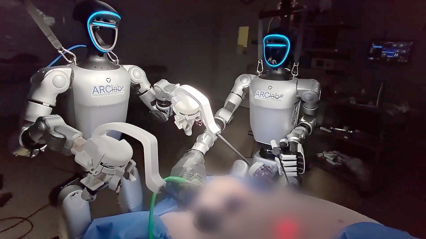
Watchers held their breath as the robot made its first incision. Hovering over its patient, an anesthetized pig, with a robotic assistant standing nearby, it navigated to the gallbladder and gently removed it.

The operation marked the debut of humanoid robots in a standard surgical setting. The robot, named Surgie, wasn’t autonomous—it was controlled by an expert surgeon—but the study is a step toward using humanoid robots as collaborators in minimally invasive surgery.

“Remotely operated and autonomous humanoid robots have real potential for amplifying access to critical surgeries to which patients would otherwise not have access,” said study author Michael Yip at UC San Diego.

The study included two successful surgeries. Human surgeons remained on standby for emergencies, but the teleoperated robot completed the task with only minimal intervention.

Feedback from surgeons operating Surgie was positive. They reported less physical strain and frustration, along with better overall performance. But they also pointed to practical problems like intermittent overheating and the need to frequently reposition the robot.

Despite a long road ahead, humanoid robots “have a viable future,” said Yip. “You can imagine these robots being deployed in remote communities where staffing is challenging, or in austere environments like search and rescue scenarios where a massive deployment of field medicine is needed in a short period of time.”

Smooth Operator

Robots have assisted surgeons for years. With a human surgeon at the helm, they excel at delicate procedures requiring precision and dexterity. They’re especially well-equipped for laparoscopic surgery, a minimally invasive technique that uses tiny incisions to reduce pain, speed recovery, and lower the risk of infection.

Despite the promise, surgeons face tradeoffs when they use surgical robots. The robots are highly specialized and often require operating rooms to be redesigned to accommodate them.

A major reason for this is the way they’re built. Intuitive Surgical’s Da Vinci system, for example, uses a robot with multiple arms, each independently controlled from a remote console. Other systems, such as Versius from CMR Surgical, deploy several lightweight independent arms, each attached to a mobile base. The robots have to be carted near the patient.

Surgeons operate all these systems from a console using a magnified, high-definition, 3D view of the surgical field, which is often better than what they’d see with their own eyes. Da Vinci 5 adds sharper visuals and depth perception with two cameras, one for each eye. And because the cameras are held by a robot rather than a human assistant, the image is far more stable.

These platforms are already used in a range of operations. But they have weaknesses. Most require proprietary surgical instruments and methods to make extra space for robot docking and maneuvering during procedures. Staff training adds further complexity and cost, limiting where the systems can be deployed.

Humanoid robots, in contrast, are far more mobile and compact. Their human-like bodies could move through standard operating rooms, use conventional surgical instruments, and potentially be easier to incorporate into existing operating rooms.

The timing may also be right. Recent advances in electric components controlling their motion have made humanoid robots faster and more stable than their awkward, stumbling predecessors. Newer AI systems that predict full-body movement and provide feedback have improved robots’ balance and ability to adjust to real-world complexities. Humanoid robots are already stocking warehouses and winning marathons.

But surgery sets a higher bar.

We still don’t know how close humanoid robots are to meeting the requirements for surgical procedures, wrote the team. That’s what they set to find out.

Hello, Surgie

The new system consists of a surgeon’s control console and the robot itself. The surgeon wears a stereoscopic headset with a magnified 3D view of the surgical field and controls the robot with an input device. The robot translates the surgeon’s commands into movements in real time.

The team chose the commercially available Unitree G1 for the job. Unlike Da Vinci, which was built for surgery, G1 is a more general-purpose humanoid with dexterous wrists and multiple joints. The researchers customized the robot’s hands so that it can rapidly switch between surgical tools. Standing just over four feet tall and weighing roughly 77 pounds, the robot takes up a fraction of the space needed by conventional surgical robots.

Precision is key for laparoscopic surgery. Surgical instruments must pivot around a fixed site at the incision, allowing them to move freely inside the body without stretching or tearing neighboring tissues. After extensively mapping Surgie’s movements, the team identified a safe set-up with enough range of motion for most minimally invasive surgeries.

Surgie passed standard robotics benchmarks evaluating surgical skill for both humans and robots. But the real challenge came next. The team performed two gallbladder removal surgeries in a standard operating room. Both operations followed a typical workflow, with a lead surgeon and an assistant responsible for placing the camera, cleaning lenses, and swapping instruments.

Surgie collaborated with the human assistant to locate, identify, and remove the gallbladder with minimal damage to surrounding tissues, including the liver. During part of one procedure, a second humanoid briefly took over camera handling while the human assistant stepped aside.

Both operations went relatively smoothly. One involved minor bleeding and bile leakage from the gallbladder, but both were easily managed. In interviews, surgeons said controlling humanoid robots felt intuitive, particularly because they had two arms and could use standard surgical tools.

“We were surprised at how well Surgie meshed with our workspace and workflow,” said study author Nikita Thareja.

The system is still in early development. Surgie’s restricted reach required frequent repositioning and recalibration, adding more than three minutes each time. The robot also occasionally needed cooling breaks after overheating. In a real operating room, interruptions like these could increase risk by forcing surgeons to split their attention between the procedure and supervising the robot.

Still, Surgie has a leg up on conventional surgical robots: It can walk. Beyond assisting with an operation, it could potentially fetch surgical tools or help clean operation rooms between procedures.

The team is now refining the system to reduce control lag, particularly during long-distance teleoperation, and exploring ways to safely sterilize—or “scrub in”—a humanoid robot for the operating room.

“Our goal is an operating theater of the future, where humanoid robots and humans work side by side as an integrated team to deliver procedures to those in need, both in traditional hospital settings as well as in non-traditional, field medicine scenarios,” said Yip.

The post In a First, a Humanoid Robot Performed Live Surgery Under a Surgeon’s Control appeared first on SingularityHub.

  •  

CAR T Revolutionized How We Treat Blood Cancers. Now It’s Closing In on Solid Tumors.

Separate teams discovered the same target in solid cancers, enabling a powerful two-pronged attack on both tumors and the cells shielding them.

Cancer researchers just found a new way to take on tumors.

CAR T cell therapy revolutionized blood cancer treatment by supercharging a patient’s own immune cells to hunt down cancers. But the approach has struggled in solid cancers. These are some of our top killers—breast, lung, prostate. Roughly two million Americans are expected to be diagnosed with cancer in 2026, and over 600,000 will likely succumb to the disease.

Unlike blood cancers, solid tumors rarely share a single, universal target for CAR T cells. Even cells within the same tumor are a mishmash. Some have little or none of a target protein, allowing them to evade the engineered immune cells, survive treatment, and fuel relapse.

“Target discovery remains a considerable challenge in the development and translation of

CAR T cell therapies for solid tumors,” wrote Christopher Mount and Marcela Maus at the Massachusetts General Brigham Cancer Institute.

Now, two independent teams have converged on the same promising target: A cell-surface protein called GPNMB. In one study, CAR T cells engineered to recognize GPNMB rapidly destroyed glioblastoma—a lethal brain cancer—in tissues taken from patients and shrank tumors in mice.

A second team used a similar strategy against an aggressive soft tissue cancer to fight tumors in organoids and mice. In an early clinical trial involving a single participant, one infusion stabilized the disease for three months without serious side effects.

CAR T designers are often wary of broadly shared targets because they can trigger dangerous attacks on healthy tissue. But GPNMB is an odd duck. In addition to cancer cells, it also sits on immune cells that spur cancer growth or suppress the body’s innate ability to get rid of tumors.

“Our approach attacks both the tumor and the environment that allows it to thrive,” said Sheila Singh at McMaster, who led the glioblastoma study, in a press release. “We’re going beyond targeting the cancer alone and eliminating the immune cells that help shield it from treatment.”

Cancer Fortress

Solid cancers have plenty of tricks to outsmart CAR T cells.

Researchers make these supercharged immune cells  by extracting a patient’s own T cells and genetically engineering them to produce protein “claws” that latch onto a specific cancer target. After infusing the cells back into the body, they seek and destroy tumor cells. CAR T has transformed treatment for several blood cancers and is showing promise in autoimmune diseases and excessive heart and kidney scarring. To simplify the procedure, researchers are also exploring ways to directly transform T cells inside the body with gene therapy.

Solid cancers, however, are far tougher opponents. Unlike blood cancers, which are heavily coated with a shared target called an antigen, solid tumors are molecular patchworks. Cells within the same tumor can display different targets—or none at all—allowing some to evade a CAR T attack and trigger relapse. Many of these targets also appear on healthy tissues, raising the risk of dangerous side effects. And then there’s the tumor microenvironment: A toxic, glue-like “fortress” that hijacks immune cells and uses them to battle incoming CAR T cells.

These barriers aren’t impenetrable. Previous work enlisted  bacteria to help CAR T cells burrow into tumors. Other efforts engineered ultra-sensitive CAR T cells capable of detecting tiny amounts of a cancer target shared across multiple solid tumors.

“Recent reports of activity in several clinical trials reinforce optimism that these efforts may result in true clinical benefit,” wrote Mount and Maus, who were not involved in either study.

But these strategies require additional engineering steps, increasing complexity and cost. And most still leave one major roadblock intact: The tumor’s immune defenses.

One-Two Punch

In the glioblastoma study, the team at McMaster University scoured donated tumors for proteins that distinguished the most aggressive cancer cells. They found one standout: GPNMB. Another test of every protein dotting the cell surface confirmed it as a promising target. The protein is evident across a cancer cell’s membrane, making it readily accessible to CAR T cells.

In lab tests, CAR T cells engineered against GPNMB performed well, nearly eliminating tumors grown from patient samples and extending survival in mice.

The target turned out to be far more valuable than expected. The team soon realized that GPNMB also marked the immune cells that suppress anti-cancer drugs. CAR T cells attacked both fronts simultaneously, weakening the tumor’s immune shield and killing the cancer itself.

“Most approaches have focused on killing cancer cells alone,” said study author Shan Grewal. “Our work suggests we may also need to dismantle the immune support system that helps the tumor survive.”

The second team focused on alveolar soft-part sarcoma, a rare soft-tissue cancer that often spreads to the lungs, brain, and bones before it’s diagnosed. Treatment often comes too late.

The disease is driven by a type of “fusion” gene created when pieces of genetic material are accidentally stitched together. These genes are extremely tough to target directly. Instead, the team screened all surface proteins on the cancer cells and again landed on GPNMB as a top candidate for intervention. The protein’s levels closely tracked the activity of the fusion gene.

CAR T cells targeting GPNMB cleared tumors and prevented metastasis in mice. But because an earlier antibody drug against the protein caused severe skin toxicity in patients, the team also tested their CAR T cells in mice carrying small human skin grafts. Although inflammation initially flared, there were no signs of ongoing skin damage.

Encouraged, the team treated a patient with relapsed, metastasized alveolar soft-part sarcoma. After a single infusion, the engineered cells rapidly divided in the bloodstream and remained detectable for roughly a month. The treatment didn’t trigger skin rashes or more dangerous side effects, like cytokine release syndrome where the body mounts a hyperactive immune defense that harms healthy organs.

The treatment’s benefits outlasted the engineered cells themselves. For roughly three months, imaging tests found fewer of the small, round spots on the patient’s lungs that often signal metastatic cancer, suggesting the disease had stabilized.

A final analysis identified another roadblock: Clusters of cells that suppress the immune system and could blunt the benefits. Adding drugs to block these immune molecules boosted tumor killing in mice. Because the same kind of gene fusion drives other cancers, including kidney, the CAR T cells could have reach beyond this specific type of sarcoma.

Together, the studies underscore that the best CAR T targets might extend beyond cancer cells to expose and attack cancer’s immune cell supporters too. Finding a viable target is a delicate balancing act. Chosen well, and CAR T cells could tackle multiple drivers for cancer growth. Choose poorly, and healthy tissues could get hurt in the crossfire.

Even so, “these two studies indicate that GPNMB represents an actionable target for CAR T cell therapies in several solid tumors,” wrote Mount and Maus.

The post CAR T Revolutionized How We Treat Blood Cancers. Now It’s Closing In on Solid Tumors. appeared first on SingularityHub.

  •  

How the Bilingual Brain Switches Languages With Ease

Similar concepts in different languages share an address in the brain.

My octogenarian father-in-law is trilingual and a lifelong fan of the World Cup. As he cheers on his favorite teams in English, Spanish, or French—sometimes switching between them mid-sentence—I’m always amazed at how easy it seems.

Scientists have long been fascinated by the brain’s ability to learn and retain multiple languages. Even after years of disuse, a brief exposure can quickly revive a language without having to consciously relearn its grammar or vocabulary. Bilingualism may offer other cognitive perks. Small studies suggest it delays brain aging, lowers dementia risk, and provides a slight edge in executive function (the ability to stay focused on a goal).

But most  of the evidence is from brain imaging studies that offer only a bird’s-eye view of neural activity and miss the finer details.

Now, scientists from the Baylor College of Medicine and collaborators have recorded activity from single neurons in four bilingual volunteers with epilepsy as they listened, read, and spoke in English and Spanish. The participants already had electrodes implanted in the hippocampus—a brain region critical for learning and memory—to track the source of their seizures.

“This is the very first study to look at how bilingual brains work at the level of individual neurons, and to do so in real time,” said study author Xinyuan Yan in a press release.

The results suggest the bilingual brain operates on two levels. Individual neurons often showed a strong preference for one language when participants heard or spoke words with the same meaning. But networks of neurons were largely language independent. They spontaneously organized into a concept map, placing words with related meanings—such as “dog” and “wolf”—closer together than unrelated words like “fork.”

Surprisingly, both languages relied on the same underlying map. Using the English concept map alone, the team could accurately predict clusters of related Spanish words.

“It’s like looking into a room from a different window. Everything inside is the same, but the perspective is different,” said study author Sameer Sheth.

Bridging Worlds

Language is central to human connection. Although some words don’t directly translate, people can express the same ideas across multiple languages without losing their core meaning.

Children raised in multilingual households are especially adept at switching between languages, often blending words and phrases together. Even when languages differ dramatically in grammar, syntax, and pronunciation, the brain somehow keeps their structures distinct while fluidly merging their meanings.

Long before we learn to speak, neural networks transform thoughts into electrical patterns that form words and sentences. Because languages are built differently—for example, where a verb falls in a sentence—it seems reasonable that each language would have a unique neural fingerprint.

But that might not be the case. A recent AI-powered analysis of functional MRI (fMRI) scans from monolingual speakers of 21 languages suggested that languages share a similar neural scaffold that represents meaning and concepts. Even fictional languages, including Klingon from Star Trek and Na’vi from Avatar, appear to tap into the same underlying system.

A growing body of evidence from bilingual speakers echoes these findings. One fMRI study found native Chinese speakers learned English more efficiently when they recruited brain networks used for Chinese. Another study identified shared speech-related brain activity sufficient for decoding words across languages.

Despite hinting at a universal language map, these standard imaging technologies struggle to capture detailed patterns as people switch languages in real time. To see how bilingual brains actually pull off the feat, we need to listen in on single cells.

Mapping It Out

The team studied four volunteers fluent in English and Spanish. All had learned the languages before age five and continued to use them regularly. Each also had electrodes implanted in the hippocampus to monitor seizures as part of epilepsy treatment, allowing researchers to track individual neuron activity as they listened and spoke.

Though often overlooked in language research, the hippocampus is increasingly recognized as a hub for word meaning, and it may also link concepts together. Here, the team monitored more than 100 neurons in each participant as they completed three language tasks.

First, the participants listened to roughly an hour of YouTube videos and the audiobook Eat Pray Love (Come Reza Ama). Next, they read aloud nearly 100 phrases displayed on a screen, such as “let’s have fun” and its Spanish equivalent “vamos a divertirnos.” Finally, they spent up to 90 minutes chatting with native speakers of each language, discussing everything from family to their epilepsy journey.

By the end, the team had compiled thousands of spoken words, hundreds of matched phrases, and hours of natural conversation.

A Language Landscape

Only a handful of neurons appeared truly bilingual, responding similarly to equivalent words such as “friends” and “amigos.” To better interpret the neural activity, the team turned to mBERT, Google’s multilingual language model that understands more than 100 languages. Like other LLMs, the model represents words according to their relationships and context rather than simple dictionary definitions.

The comparison revealed a similar pattern in brains and machines. Individual neurons rarely encoded the same word across languages. Instead, meaning emerged at the population level.

Both neural activity and mBERT tracked broader context, organizing words into an abstract conceptual landscape called semantic geometry. In this map, related concepts cluster together—“cat” sits closer to “dog” than to “galaxy,” for example—even if the precise features defining those relationships are unclear.

Yet the map remained largely unchanged across languages, suggesting it captured a fundamental mechanism for language processing in the brain. Using the English map alone, the team could predict which Spanish words would cluster around “perro” (or “dog”).

“This is how the brain encodes the meaning of words across languages,” said Yan. “It doesn’t rely on individual neurons translating individual words, but groups of neurons adjusting their activities to create the similar pattern for equivalent words in both languages.”

The study focused on semantics, or meaning, as opposed to syntax, the rules governing sentence structure. A recent study also using single-cell recordings from people with epilepsy suggests that other groups of neurons, particularly those in the frontal parts of the brain, may specialize in grammar while ignoring semantics. Whether they also share a “map” across languages remains to be seen.

The next step is to watch these maps emerge. The team hopes to track people as they learn a new language, revealing how new words and concepts are woven into semantic landscapes in real time. The results could deepen our understanding of one of the most fundamental communication skills and even inspire more capable and efficient language models in AI.

“Our study shows that the brain is wired to learn multiple languages,” said study author Benjamin Hayden.

The post How the Bilingual Brain Switches Languages With Ease appeared first on SingularityHub.

  •  

Woman With Alzheimer’s Shows Striking Improvement After Taking Magic Mushrooms

A single observational case suggests psilocybin may ‘awaken’ cognitive reserve in dementia. But scientists caution controlled trials are needed to know if the drug was the cause.

For five years, Alzheimer’s slowly stripped away a Japanese-American woman’s ability to speak more than one syllable at a time. The woman, now in her 80s, was diagnosed roughly a decade ago, and her condition steadily worsened. She struggled to walk and recognize family members.

Then, under medical supervision, she took a large dose of mushrooms containing the psychedelic psilocybin. Within three days, her symptoms had improved. She began spontaneously recounting memories and initiating conversations in full sentences. Her alertness returned, and she could move around independently.

A week later, she was recognizing family members, asking where they were, and pointing out cars that seem out of place.

Psilocybin has been maligned for decades. But renewed interest in its unique effects on the brain has pushed it into mainstream research. Early studies suggest it may help treat depression, anxiety, addiction, post-traumatic stress disorder, and other psychiatric conditions. A clinical trial is underway to gauge whether it can protect the aging brain.

The case study, conducted in Brazil, adds to that momentum. The team emphasizes that it describes a single patient and is purely observational. Because of the severity of her disease, they could not perform brain scans, measure biomarkers, or conduct standard cognitive tests. Exactly why her symptoms improved remains unknown.

Even so, they propose that psilocybin may have temporarily unlocked brain function in late-stage Alzheimer’s, potentially allowing dormant neural networks to rewire.

Brain Under Fire

Alzheimer’s is often synonymous with memory loss. Sadly, symptoms range far beyond forgetting names or misplacing glasses.

As the disease progresses, people gradually struggle to find the right words or follow conversations. Their ability to tackle everyday tasks—cooking, managing finances, planning ahead—erodes. Depression, irritability, and anxiety often emerge. Over time, their personalities flatten, leaving them less outgoing, engaged, or empathetic.

These stories are far too common. According to the World Health Organization, roughly 57 million people worldwide were living with dementia in 2021. Alzheimer’s may account for up to 70 percent of cases. As populations age, that number is expected to climb.

Alzheimer’s has no single cause. Genetics likely play a role. Some gene variants are linked to early-onset forms of the disease, an area scientists are now tackling with gene therapy.

Another hallmark of the disease is a buildup of abnormal protein clumps, or plaques, in and around neurons, which disrupts normal function and wrecks their ability to form neural networks supporting memory and cognition. Years of efforts to remove plaques have largely failed, though the FDA recently approved two antibodies that reduce them and modestly slow cognitive decline.

Then there’s inflammation. In Alzheimer’s, the brain’s immune system can become overactive. Rather than responding only to damage, inflammation drives disease progression, spreading toxic protein clumps through the brain and further damaging its ability to form new connections.

Here’s where psilocybin, the active ingredient in magic mushrooms, may help. Psilocybin alters serotonin signaling, a brain chemical involved in mood, perception, and cognition. But its effects likely extend far beyond that.

Studies in mice suggest the chemical boosts the brain’s ability to rewire, a process known as neuroplasticity. Human brain imaging studies have found that the psychedelic temporarily reorganizes communication between large brain networks, changing how distant regions interact. In some participants, supervised treatment has been linked to greater cognitive flexibility, deeper self-reflection, and improved well-being.

Other studies hint at a protective role. Psilocybin triggers the release of “nurturing” proteins. This process helps neurons survive stress and extend their branching connections. It’s these delicate structures that build up neural networks, and they wither away during depression, aging, and dementia. Inside the hippocampus, a region crucial for learning and memory, the drug stimulates the birth of new neurons, at least in mice.

Given its positive effects on brain plasticity, psilocybin is now being tested in multiple psychiatric disorders characterized by unusually rigid patterns of brain activity. Older adults remain largely absent from these studies, even though they could benefit the most.

Tale of One

Before treatment, the woman struggled with everyday life. For five years, she could communicate using only single-syllable words. Her mobility was severely limited, and she struggled with incontinence.

With the consent of her caretaker, she received five grams of the Enigma strain of Psilocybe cubensis. Because psilocybin levels vary widely between mushrooms, the exact dose is unknown. But compared to other clinical trials, it was relatively high.

The team chose the dose “based on prior experiential observations regarding depth and duration of psychedelic-induced neurobehavioral effects,” wrote the team.

Initially, the woman fell into a deep sleep-like state accompanied by elevated body temperature and heavy sweating. Roughly 19 hours later, she suddenly awoke and began speaking to caregivers in complete sentences, recounting memories from her life. The conversation lasted around four hours.

Over the following days, she became increasingly alert and engaged. She recognized family members, regained mobility, and could pick out matching clothes to dress herself. A week later, she was noticing small details in her environment, including a rental car parked outside the house. When a family member was absent, she asked, “Where did Celso go?” She also seemed to rediscover her love of social interactions, making eye contact, smiling back, and actively starting conversations.

A month after the initial session, she returned for a second supervised dose of three grams. After the second dose, she became even more verbally expressive, displayed a sense of humor, and described memories of surfing with her son on a peaceful island. Throughout the trial, the drug alleviated incontinence and improved her quality of life.

The results come with major caveats. The improvements were observational and largely reported by caregivers, leaving room for bias. The team didn’t administer standardized tests for cognition, dementia, depression, and anxiety. Nor did they perform brain scans or monitor sleep, making it impossible to determine what brain changes were behind her apparent “awakening.”

“Causality cannot be established, and spontaneous fluctuations inherent to neurodegenerative disease cannot be completely excluded,” they wrote.

But the study touches on a provocative idea in Alzheimer’s: Cognitive reserve. The theory proposes some people can tolerate greater levels of harm to the brain and continue functioning despite significant damage. Psilocybin may have temporarily tapped into these reserves, allowing dormant neural circuits to engage and rewire to compensate for impaired ones. The hypothesis is highly speculative and needs to be rigorously tested.

Meanwhile, a clinical trial is investigating whether psilocybin can reduce depression and improve quality of life in people with mild cognitive impairment or early Alzheimer’s disease, moving the needle beyond a single case study.

For one family, however, the benefits are already substantial. At a follow-up visit, the woman spontaneously said to everyone in the room, “It is pleasant to come here.”

The post Woman With Alzheimer’s Shows Striking Improvement After Taking Magic Mushrooms appeared first on SingularityHub.
  •  

AI Collapses on a Classic Psychology Test. What It Reveals Could Stall Human-Level AI.

AI needs to focus more like we do.

“Attention is all you need.”

This 2017 breakthrough idea transformed AI. The concept of self-attention became the foundation of today’s chatbots. Claude, Gemini, and ChatGPT are all large language models (LLMs), AI systems designed to focus on the matter at hand while filtering out distractions.

The results have been remarkable. From brainstorming recipes to generating code, apps, websites, and content, LLMs are being woven into our lives at breakneck speed.

But now, a City University of New York team and collaborators are asking: How closely does AI self-attention resemble human attention?

It’s not just academic curiosity. AI researchers have long looked to the brain for ideas to improve machine intelligence. In turn, AI models have offered new ways to investigate the brain. Comparing artificial and biological attention could inspire AI that concentrates more like us.

In their study, the team asked multiple chatbots to complete a classic psychology test of attention and cognitive control. Participants are shown the word for a color—such as “red”—written in either the same or a different color than the one the word describes. The challenge is to name the ink color while ignoring the word itself.

On short word lists, the chatbots performed at a high level. But as the tasks grew longer, their focus faltered. Instead of naming the ink color, they increasingly defaulted to reading the word. Under more demanding conditions—ones that also trip up people—their performance nearly collapsed.

The findings suggest today’s AI attention systems are “fundamentally limited,” wrote the authors. They go on to say that adding mechanisms similar to “those in biological attention is crucial for achieving artificial general intelligence.”

Attention, Two Ways

Doomscrolling. YouTube. Dinner plans. Family obligations. A barrage of notifications.

Life sometimes seems like everything, everywhere, all at once. Yet the brain can usually lock onto what matters most and push everything else into the background.

Far from a single, straightforward mechanism, attention emerges from multiple brain regions. According to attention network theory, three networks do most of the heavy lifting.

The alerting network keeps the brain ready for action. The orienting network selects which sights, sounds, smells, and sensations deserve attention. Finally, the executive control network resolves conflicts between competing streams of information, helping direct thoughts and actions toward a goal.

Together, these systems allocate the brain’s limited resources. Touch a hot stove, for example, and your brain immediately shifts attention to the burn over dinner. The food can wait; cooling your hand can’t.

AI works very differently.

Rather than processing language as complete sentences, LLMs break text into smaller units called “tokens.” Attention mechanisms then determine which tokens matter most for generating the next word, sentence, or response.

Self-attention is the key breakthrough behind modern chatbots. For each token, the model weighs and incorporates information from other tokens in a sequence, allowing it to track context across long stretches of text. This mechanism helps AI connect words and ideas, and underpins virtually all frontier LLMs today.

Researchers have since built on the concept. One approach, multi-head attention, runs several attention systems in parallel, with each “head” learning different patterns, such as grammar, syntax, or meaning. Another, cross attention, links information across different chunks of inputs and their outputs, making it especially useful for tasks such as translation and summarization.

But attention comes at a steep computational cost. To make models more efficient, researchers are also exploring sparse attention, which limits how many tokens a model considers at once. Another approach draws on information learned in the past to keep AI “focused.”

Despite the name, AI attention is ultimately a mathematical system. It helps determine what information is relevant in a specific context. But it lacks executive control, the network that keeps humans continuously focused on a goal despite distractions for long periods of time.

Color Blind

To test the limits of AI attention, the team pitted OpenAI’s GPT-4o and Anthropic’s Claude 3.5 Sonnet against the Stroop task.

Invented by John Ridley Stroop in 1935, the test measures attention and cognitive control by forcing participants to resolve conflicting information. The challenge is simple: Name the color of a word while ignoring what the word means. In a congruent trial, the word “blue” appears in blue ink. In an incongruent trial, “blue” might appear in red or green, creating a conflict between what the eyes see and what the brain reads.

Humans are consistently slowed down by this interference. Even with practice, the effect remains, suggesting it taps into fundamental mechanisms of executive control.

In the study, the researchers created word lists of varying lengths and difficulty. Some were entirely congruent. Others were fully incongruent. A third set mixed the two conditions.

At first, the AI models excelled. On five-word tests, GPT-4o was over 90 percent accurate across all conditions. But as the number of words increased, performance plummeted. On 40-word incongruent tests, the model’s accuracy fell to roughly 15 percent. Claude showed a similar decline. In mixed-condition tests, both models’ performance nearly collapsed to zero.

“The sharp decline in color-naming accuracy with increasing list length indicates that transformer-based attention mechanisms are vulnerable to scaling demands,” wrote the team.

Perhaps most intriguing, some models correctly recognized they were taking the Stroop test and could even explain its rules. But that apparent awareness did nothing to improve their scores. In other words, a “book smart” understanding of the task wasn’t enough to execute it well.

The study joins a growing effort to borrow psychological tests for research in machine cognition, especially when AI is challenged with complex, dynamic decision-making tasks. Theory of mind tests, for example, let researchers gauge whether a system can track others’ beliefs, emotions, and intentions. Personality tests are helping shape model behavior and reduce sycophancy. And some LLMs are readily solving emotional intelligence tests, which measure how well the algorithms recognize and respond to social cues.

According to the authors, the new results point to a missing ingredient in AI attention: A mechanism similar to the brain’s executive control network, which helps us stick to a task and adapt when priorities change.

Future AI systems could benefit from higher-level executive control that continuously tracks progress toward a goal, detects when attention has drifted, and pulls it back on course, if necessary.

Rather than simply weighing which tokens are most relevant in the moment, a more human-like form of attention could help AI stay focused during complex tasks, such as long conversations, multi-step reasoning problems, or high-stakes use in scientific research and drug discovery.

“The ultimate goal of AI research is to develop artificial general intelligence comparable to human abilities,” wrote the team. “AI systems, like humans, may need to master fundamental attention mechanisms…before achieving the generalized problem-solving abilities characteristic of mature executive functions.”

The post AI Collapses on a Classic Psychology Test. What It Reveals Could Stall Human-Level AI. appeared first on SingularityHub.

  •  

Can Psychedelics Reboot Aging Brains? We’re About to Find Out

An audacious trial will test psilocybin in people over age sixty to see if increases plasticity in healthy aging brains.

A handful of healthy senior citizens are about to trip on psilocybin—to see if the psychedelic protects aging brains.

Psilocybin, the active ingredient in magic mushrooms, is best known for its ties to 1960s counterculture. But now it may also herald a new genre of mental health treatment. From severe depression to post-traumatic stress disorder, studies have highlighted psychedelic drugs’ ability to reshape brain networks and relieve debilitating symptoms.

Most of these studies have focused on younger people with mental health conditions that don’t respond to standard treatment. The field’s success is prompting scientists to ask if psychedelics could also help healthy brains age better.

A team from the UC Berkeley Center for the Science of Psychedelics is about to find out. In a first-of-its-kind study focused on adults between the ages of 60 and 85, they’ll investigate how psilocybin affects perception, emotion, and memory using a battery of psychological tests.

Multiple scans before and after dosing will track changes in the brain. And detailed surveys will gauge broader shifts in well-being: Do participants feel more “in tune” with their emotions, feel less isolated, or experience a renewed sense of wonder about the world?

“What really excites me is that we’re focused on healthy older adults,”  said Tyler Toueg, who co-led the study’s design, in a press release. “Most clinical trials with older adults are focused on people who already have a diagnosis. We’re asking whether we can actually promote positive outcomes in older adults who are healthy.”

Called PLASTICITY, the trial could also open a rare window into how a psychedelic experience reshapes healthy brain networks. And because the drug alters our sense of self, psilocybin could help researchers probe the ways in which the brain constructs reality.

“I’m very interested in psilocybin as a potential mental health treatment, but I’m also interested in it as a way to shed light on these central mysteries in neuroscience and psychology,” said study designer Michael Silver.

Chasing the White Rabbit

Psychedelic research was highly restricted for decades. But advocates, including the non-profit Multidisciplinary Association for Psychedelic Studies, have steadily pushed to reopen the field, arguing that these drugs might keep mental health symptoms at bay.

Early results helped usher psychedelics into the mainstream. In 2023, a randomized, placebo-controlled trial found that a single dose of psilocybin, paired with therapy, eased depression. Oregon later approved supervised psilocybin therapy—though the drug remains federally illegal in the US—and Australia became the first country to greenlight it for depression and post-traumatic stress disorder. More recently, two late-stage studies reported strong effects in severe depression, potentially paving the way for FDA approval.

Scientists still don’t fully understand how psilocybin works in the brain. But there are hints. The drug appears to rapidly reorganize the connections between brain cells, particularly in the hippocampus, a region of the brain central to learning, memory, and navigation.

Neurons constantly change their connections in a process called plasticity that encodes experiences into neural networks, allowing the brain to process information, learn, and lock in memories. In youth, these connections are flexible and expansive. But with age and in conditions like depression, the brain’s flexibility wanes.

The birth of new neurons, or neurogenesis, also contributes to plasticity. Neurogenesis only occurs in two brain regions, one of which is the hippocampus. Although whether it actually takes place in humans is controversial, it is strongly linked to learning, memory, and emotion, and it declines in both psychiatric disorders and aging.

Psilocybin may reset brain plasticity to a more youthful state.

In rats modeling depression, for example, a study showed the drug shifted dark moods into behavior that was more exploratory and engaging. Where traditional antidepressants, like Prozac, tend to blunt symptoms, psilocybin seems to overwrite entrenched negative patterns. This suggests deeper circuit-level changes.

In another study, the drug reopened a critical window for learning in mice. During adolescence, the brain is highly plastic, but it begins to stiffen in adulthood. Psilocybin temporarily restored malleability and changed the mice’s social behavior. In some brain regions, the drug increased sensitivity to oxytocin, the so-called “love hormone.” The authors suggested the drug induced a state called metaplasticity, in which neurons are more likely to respond to oxytocin and other regulators to rewire, form new connections, and grow their branches.

Psilocybin’s effects may extend beyond the brain. Depression, chronic stress, and the immune system are tightly linked. In a third study, researchers identified a brain-spleen connection that drives fear and anxiety. Psilocybin suppressed inflammatory immune cells associated with brain inflammation and dampened anxiety-like behavior in stressed mice, even under threat.

Both effects—dialing up plasticity and lowering inflammation—make psilocybin an intriguing way to potentially counter changes in the aging brain.

“We know that with age, we lose synaptic connections, especially in certain brain regions like the hippocampus and prefrontal cortex,” said Toueg. “There’s a lot of overlap between the mental states that psychedelics influence and those associated with successful aging.”

Tomorrow Never Knows

The PLASTICITY trial is designed to test whether psilocybin can produce lasting changes in neuroplasticity in healthy adults aged 60 to 85. Participants will first undergo assessments of cognition, visual perception, and brain structure using advanced MRI techniques.

Diffusion MRI scans will focus on the hippocampus to capture microscopic changes in its structure. Functional MRI will focus on brain activity as participants perform learning and memory tasks, offering a dynamic view of how activity shifts after dosing.

The study will also see if psilocybin increases vagus nerve activity, which has been linked to better stress recovery. Participants will complete detailed surveys about the experience ranging from emotional responses, like wonder, to potential shifts in outlook and social cognition.

“Things like depression, anxiety, stress and rumination are all associated with worse aging outcomes,” said Toueg. “Things like having purpose in life, emotional regulation, and awe are all associated with more successful aging.”

The trial began enrolling participants in November last year. Two volunteers have already completed the tests, and the team aims to dose 20 people by the end of 2026.

Although psilocybin trials are now widespread, older adults remain underrepresented. One estimate suggests just 1.4 percent of participants are 65 or older, despite potentially being among those most likely to benefit from interventions that enhance plasticity.

“This study allows us to directly test whether the promising findings from animal models translate to older humans and to generate data that will inform future research on aging, cognition, and mental health,” said Silver.

Toueg agrees. “I think that no matter what we find, this study will have implications for how we think about intervening in the aging brain,” he said.

The post Can Psychedelics Reboot Aging Brains? We’re About to Find Out appeared first on SingularityHub.

  •  
❌